What to expect During Examination
1. Physical Examination
Physical examination is the initial and crucial step for our dentist to assessyour oral health. It involves a thorough inspection of your teeth, gums, tongue, throat, and surrounding tissues using their trained eyes and sometimes with the aid of specialized tools.
By performing a thorough visual examination, the dentist can gain valuable insights into your oral health status. This information, combined with other aspects of the dental exam like X-rays, helps them create a personalized treatment plan to address any existing issues or prevent future problems.
2. Radiological Examination
In dentistry, a radiological examination, often referred to as dental X-rays, plays a vital role in uncovering hidden issues that a visual examination alone might miss.
These X-rays provide a deeper look into the structures beneath the surface of your teeth and jaws, aiding dentists in early detection, diagnosis, and treatment planning.
Types Of Xrays:
1. OPG X-ray
An orthopantomogram (OPG), also known as a panoramic X-ray, is a wide-angle dental X-ray that captures the entire upper and lower jaw, all your teeth (including those that haven't erupted yet), and the surrounding structures in a single image.
OPGs provide a more comprehensive view of your mouth this broader view allows dentists to
detect hidden cavities, assess jawbone health, evaluate wisdom teeth and plan dental procedures .
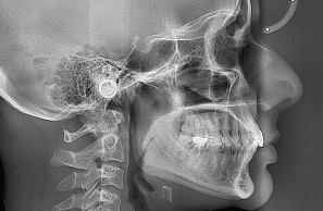
2. Lateral Cephalogram
A lateral cephalogram, also known as a lateral cephalometric X-ray, is a specialized X-ray of the head taken from the side. It captures a profile view of your skull, jaw, teeth, and soft tissues in a single image.
Lateral cephalogram provides a detailed view of the facial profile, jawbone structure, the size, tooth position, sinuses and airway.
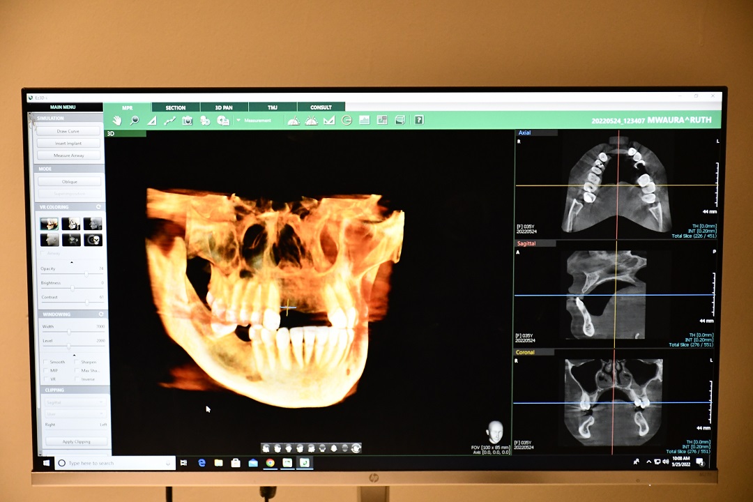
3. CBCT
Cone-beam computed tomography (CBCT) is a special type of X-ray imaging technology used in dentistry that provides highly detailed 3D images of your teeth, jawbone, and surrounding facial structures.
CBCT scan creates a three-dimensional picture by rotating an X-ray source around your head. This allows dentists to examine your oral structures in much greater detail and from different angles.
2. IOPA
IOPA stands for Intraoral Periapical Radiograph. It's a specific type of dental X-ray that captures a detailed image of one or two teeth, including the crown (visible portion of the tooth) and the root beneath the gum line. Unlike panoramic X-rays that provide a broad view of your entire mouth, IOPAs offer a magnified view of a specific tooth area.
IOPA X-rays are a safe and effective diagnostic tool in dentistry. The amount of radiation used is minimal, and dentists take precautions to minimize exposure using lead aprons and collimators